Indian Journal of Physiology
and Pharmacology
and Pharmacology
Indian Journal of Physiology and Pharmacology |
 (252x320).jpg) |
Volume 58 - Number 2 April - 2014 (Current issue) ISSN 0019-5499 |
Ameliorating Effect of Black Tea Extract on Cadmium Chloride-induced Alteration of Serum Lipid Profile and Liver Histopathology in RatsVenkappa S Mantur, Manjunath S Somannavar, Saeed Yendigeri, Kusal K Das and Shivaprasad S Goudar |
Cadmium is one among the most environmental pollutants that affects many organs like kidney, liver and testis. The present study was aimed to assess the simultaneous effects of black tea extracts (BTE) on cadmium chloride induced alterations in lipid profile and liver histology. Adult rats were divided into four groups (n=6/group), group I (normal saline), group II (CdCl2 , 1.0 mg/kg, b.wt; i.p), group III (black tea extract, 2.5 gm tea leaf/dl of water that is 2.5% of aqueous BTE) and group IV (cadmium chloride + BTE). Cadmium chloride intoxicated rats showed significant increase in serum total cholesterol, triglycerides, and low density lipoprotein-cholesterol and there is a significant decrease in the serum high density lipoproteincholesterol. In the liver, cadmium chloride showed changes in normal architecture, swollen hepatocytes, kupffer cells hyperplasia, dilation and congestion of central vein. Oral administration of black tea extracts with cadmium chloride significantly improves lipid profile and liver architecture as compared to the cadmium chloride group.The results indicate that BTE is beneficial in preventing cadmium-induced lipid alterations and hepatocellular damage. |
Metals are persistent environmental contaminants. Many metals such as cadmium, lead, arsenic etc.
have the potential to cause genetic alterations in the
target tissues of exposed humans. If they affect tumor
suppressor genes, it may lead to the development of
cancer in the target organs (1). Cadmium mainly
discharges from the battery manufactures, plastics,
fertilizers, electroplating, pesticides etc. The liver is
the main target organ when considering Cd-induced
toxicity because Cd primarily accumulates in the
liver (2). Cadmium is known to be carcinogenic, and studies have been linked with cancers in the lungs
and prostate (3). Cadmium (Cd) is present in the
soil, water, air, food and cigarette smoke. It causes
poisoning in various tissues of animals, and humans
(4). Studies have suggested that generation of
reactive oxygen species (ROS) and its interference
with cellular antioxidant system is one of the major
mechanisms by which toxic effects of cadmium is
mediated (5). The flavonoids compounds in tea are
considered as important therapeutic substances that
act as antioxidants. Moderate intake of black tea
improves the levels of independent risk factors of
cardiovascular disease and antioxidant defenses in
plasma (7). As heavy metals have been known to
possess many adverse health affects for a long time
the understanding of heavy metal toxicities and
fighting heavy metal toxicities with any chelating
agent or phytochelators or any type of naturally
available antioxidants is the need of the hour. Hence
present work is aimed to assess ameliorating effect
of black tea extract on cadmium chloride induced
alteration of serum lipid profile and liver histopathology
in rats. |
Wister strain of albino male rats aged 50 to 60 days and 160+10 g body weight was used in this study. All animals were fed with water and food ad libitum. Experiments were carried out after obtaining institutional animal ethical committee approval. Animal breeding and maintenance were done in accordance with guidelines of Government of India for use of laboratory animals (8). Preparation of 2.5% Aqueous BTE: The black tea (Camellia sinensis) extract will be prepared from CTC (Cut, Tear and Crush) BOP (Broken Orange Pickoe) grade black clonal tea. It was processed and supplied by Tocklai Experimental Station, Jorthat, Assam. A fresh 2.5% aqueous BTE was prepared every day following the method of Wei et al. (1999). Twenty five gram of black tea was added to 500mL of boiling water and was steeped for 15 minutes. The infusion will be cooled to room temperature and then filtered. The tea leaves were extracted a second time with 500 mL of boiling water and filtered, and the two filtrates were combined to obtain a 2.5% aqueous black tea extract (2.5 g of tea leaf/100 mL water). The resulting clear solution is similar to tea brews consumed by humans (10). All the animals were acclimatized for 7 days to the
laboratory conditions at 22-24°C and a 12 hr light:
dark cycle. The Wister strain male albino rat (160-180
gm) were chosen and they were divided into 4
groups i.e. group-I, control; group-II, cadmium
chloride (1 mg/kg B.Wt, I.P for 21 days) (Souad H
2010.); group-III (black tea extract dose of 2.5 gm
tea leaf/dl of water that is 2.5% of aqueous BTE
also for 21 days) and group IV (both cadmium
chloride and BTE simultaneously). All the animals
were sacrificed at the end of the last dose after
an overnight fast. Blood samples were collected
in a centrifuge tube and allowed to form serum.
Serum total cholesterol, LDL-cholesterol, high-density
lipoprotein (HDL) cholesterol, and triglycerides
were assayed using an enzymatic estimation All the experimental procedures followed were performed in accordance with the approval of the Institutional Animal Ethics Committee (1169/ac/08/ CPCSEA) under strict compliance of Committee for the Purpose of Control and Supervision of Experiments on Animals (CPCSEA) guidelines for the experimental studies. For the evaluation of histopathology, liver tissue from each rat was stored in 10% neutral buffered formalin solution. Three lobes of each rat liver were processed and sectioned. By following routine histological techniques, the samples were put into paraffin, and serial sections of 5 mm were taken from tissue blocks. For each rat, three slides were made and stained with haematoxylin and eosin. The preparations were evaluated under a photomicroscope and photo-graphed (Olympus BH-2 with Samsung Digital Color Camera, Model No. SDC242. The following changes of the tissues of control and various treated groups were observed; viz. atrophy degeneration, necrosis, inflammation, tumorogenecity etc. Statistical analysis Data were expressed as mean+standard deviation of mean (SD). Statistical comparisons were performed by one-way ANOVA followed by post-hoc t test and the values were considered statistically significant when P<0.05. |
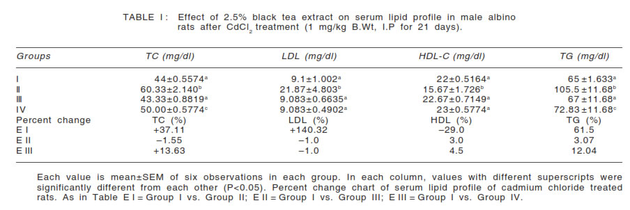
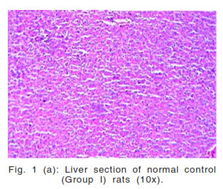
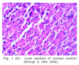
Table I shows in Group II, cadmium induced a significant increase in the serum total cholesterol, LDL cholesterol, triglyceride levels and significant decrease in HDL cholesterol level comparison with the control group I. Group IV (cadmium + BTE) also show the significantly increase in total cholesterol, triglyceride in comparison with the group I but when these values were compared with group II (cadmium alone treated), significant lower total cholesterol, triglyceride values were noticed. Table I also shows the percent change increase of Total cholesterol, LDL-cholesterol, and triglyceride and the percent change decrease of serum HDL-cholesterol in group IV in comparison with the control group I. But when compared with group II receiving cadmium chloride only, the rise in total cholesterol, LDL-cholesterol, triglyceride, and fall in serum HDL-cholesterol were remarkably less (E I vs. E III). Rats receiving BTE alone (group III) did not show any significant variation in the parameters studied above when compared with control (group I). The histological structure of normal untreated rat liver
(group I) showed a normal architecture (Fig. 1(a),
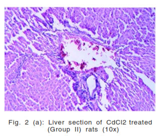
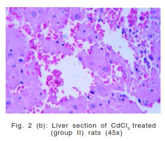
Fig. 1(b)). In group II (cadmium chloride) showed
hepatocytes appear to be swollen, dilatation and
congestion of central vein, kupffer cell hyperplasia,
distorted 'lobular' architecture of liver parenchyma also seen (Fig. 2 (b), Fig. 2 (b)), In group III (BTE)
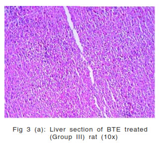
showed normal architecture, hepatocytes appear
normal, central vein appears normal (Fig. 3 (a), Fig.
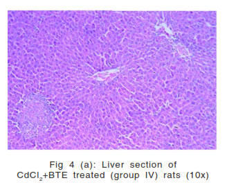
(b)). In group IV hepatocytes appear to be little
swollen, mild widening of sinusoidal spaces, mild
Distortion of 'Lobular' architecture of liver parenchyma
(Fig. 4 (a), Fig. 4 (b)). |
 |
 |
 |
 |
 |
 |
 |
 |
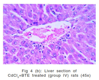 |
Our results on cadmium treatment in rats clearly exhibit hepato-toxicity by altering serum lipid profile along with liver histopathology. Increase cadmium burden in the body, increases the risk of dyslipidemia, mainly due to the increased risk of low HDL cholesterol and the high ratio of triglycerides to HDL cholesterol (12). Cadmium chloride induces increase serum total cholesterol, LDL-cholesterol and triglycerides and fall of serum HDL-cholesterol (group II rats) may be due to changes in the gene expression of hepatic enzymes like HMG-CoA reductase (hydroxy-methylglut-aryl-CoA reductase), which in turn depresses LDL-receptor gene expression (13). In the present study the significant improvement in lipid profiles in group IV rats treated concomitantly with BTE and cadmium chloride agrees with reports that BTE can protect against the serum and hepatic abnormalities in hypercholesterolemic rats (14). This report was also in agreement with the observation that total plasma lipid peroxides and oxidation of LDL were significantly reduced in hamsters fed a high cholesterol diet and given black tea (15). It was further observed that five times daily serving of black tea reduced low density lipoprotein (LDL) by 11.1% and total cholesterol by 6.5% in hypercholesterolemic adults (16). No change of lipid profile in group III rats after BTE supplementation alone as compared to control (group I) may be due to abilities of BTE to keep all the lipid profile parameters within normal range. The liver is an important organ performing vital functions including biotransformation, migration of lipids glycogen storage and release of glucose into the blood. The results of our histopathological studies show hepatocytes appear to be swollen, dilatation and congestion of central vein, kupffer cell hyperplasia, distorted 'lobular' architecture of liver parenchyma also seen (Fig. 2(a), Fig. 2(b)). The histopathological studies in the liver of rats treated with cadmium clearly indicated an extensive liver parenchymal damage like swollen hepatocytes, congested and dilated central vein and kupffer cell hyperplasia [Fig. 2(a), Fig. 2(b)]. These histopathological observations clearly indicate that cadmium is hepatotoxic. The change in cellular integrity could be due to the oxidative stress developed by the nickel-induced generation of reactive oxygen species (ROS). There is significant improvement in the histopathology of the liver in rats treated with cadmium chloride and BTE (Fig. 4 (a), Fig. 4 (b)). Hepatocytes appear to be little swollen, mild distortion of 'Lobular' architecture of liver parenchyma, mild widening of sinusoidal spaces observed in BTE + cadmium treated group (Fig. 4 (a), Fig. 4 (b)). Mild distortion of lobular architecture of liver parenchyma, with widening of sinusoidal spaces was observed in BTE alone treated group [Fig. 3 (a), Fig. 3 (b)]. Hence it may be concluded that cadmium chloride is a toxic heavy metal, that-affects the serum lipid profile and causes degenerative histopathological changes in rat liver. Simultaneous treatment with BTE partially improved cadmium chloride induced hepatocellular damage and serum lipid profiles. Limitations The limitation of this study is as authors have not estimated some active compounds like theophylline or theobromine in the tea extract hence it is difficult to conclude which chemical in the black tea restores lipid profile following injections along with cadmium. |
|